Scientists at IBM Research in Zurich, Switzerland, have made a humongous breakthrough involving one of nature’s smallest building blocks: the molecule.
A team of IBM Zurich scientists on Monday announced they have produced the first-ever image of how an electrical charge is distributed throughout a single organic molecule called naphthalocyanine — only two nanometers long (that’s two billionths of a meter).
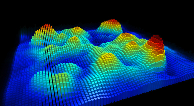
Actually, make that several images that the IBM team has cooked up: The team posted a series of its work on Flickr, including black-and-white models and artificially colorized ones for clarity’s sake, even a 3D map of the charges generated from the image data (the picture included at the top of this article).

Right now, the imagery is mostly a testament to the increasing powers of modern, nano-scale microscopes. IBM notes that the imagery does provide a clearer portrait of the composition of molecules, comparing the technique to giving someone an X-ray to find out what’s going on inside their bodies.
But in the future, IBM hopes that it will be able to get images of molecules undergoing changes in the distribution of their electrical charges when forming chemical bonds and interacting with other substances.
Ultimately, the technology could be used to produce even smaller computer chips, higher-efficiency solar cells and better batteries.
“In particular, the technique could contribute to the design of molecular-sized transistors that enable more energy efficient computing devices ranging from sensors to mobile phones to supercomputers,” said Chris Sciacca, an IBM spokesperson in Zurich, in an email to TPM.
IBM published the news of its latest advancement in the journal Nature Nanotechnology late Sunday.
The Zurich researchers managed to obtain the fantastic, ultra-close through a technique known as Kelvin probe force microscopy (KPFM). This technique involves taking a microscope with an extremely fine, nano-size tip and putting it above an electrically-conductive molecule, then switching on a power current.
Electrical fields are generated between the tip and the molecule, and the microscope measures the resulting fields to determine the rough shape of the molecule’s charge distribution.
The fields are stronger above areas of the molecule that carry the electrical charge, and weaker over those that don’t, producing an image where the brighter colors correspond to stronger charges, the more faded tones to weaker charges and the deep, dark tones to areas of the molecule that are oppositely charged. The actual charge itself is not visible.
“To achieve submolecular resolution, a high degree of thermal and mechanical stability and atomic precision of the instrument was required over the course of the experiment, which lasted several days,” Sciaccia told TPM.
IBM chose naphthalocyanine, a molecule comprised of two hydrogen atoms opposite each other in the center, because it had some previous experience with it, using naphthalocyanine to create the world’s smallest logic switch (the basic “on and off” command at the heart of all computational devices) in 2006.
But it will likely be a few years before it can truly harness the power of its most recent discovery. That hasn’t stopped scientists around the world from applauding IBM, with one scientist, University of California Berkeley’s Michael Crommie, telling Computerworld that he thought the imaging could be used to create more effective graphene — the strongest, thinnest nanomaterial yet synthesized.


